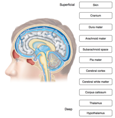
Place the following items associated with the brain in order from superficial to deep.
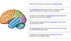
Complete each sentence describing the structures and functions of the cerebrum.
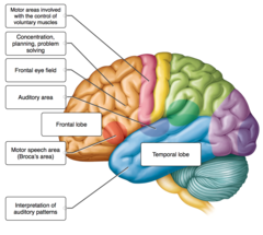
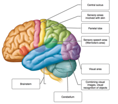
Consider a situation in which a stroke or mechanical trauma has occurred, resulting in damage one of the areas of the brain indicated in the image. Drag and drop each label, identifying the cerebral area that, if injured, would result in the functional disturbance described.
The labels list functions of various areas of the cerebrum. Place each label on the appropriate cerebral lobe.
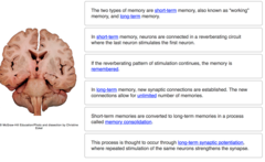
Complete the following sentences describing various aspects of memory.
The limbic system interprets sensory impulses from the receptors associated with which sense?
smell
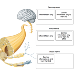
Determine whether the following descriptions apply to sensory nerves, motor nerves, or mixed nerves (both sensory and motor).
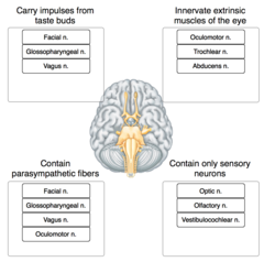
Place each cranial nerve label in the appropriate category, describing its function.
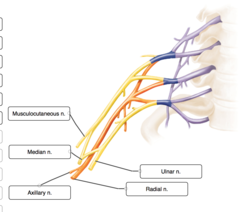
Label the terminal branches of the brachial plexus.
Label the structures of the central nervous system and their protective structures.
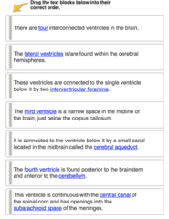
Complete the sentences describing the ventricles and spaces associated with CSF in the central nervous system.
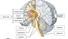
Label the internal structures of the cerebrum and other major parts of the brain.
Label the indicated lobes of the cerebrum and the functional areas of the cerebral cortex.
Label the indicated brain structures, cerebral lobes, and functional areas of the cerebral cortex.
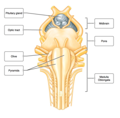
Label the anterior view of the brainstem.
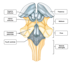
Label the posterior view of the brainstem.
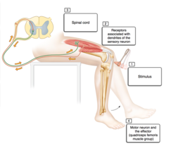
Place in order the structures and/or events associated with the patellar reflex.
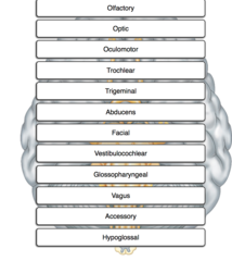
Place cranial nerves in numerical order, beginning with cranial nerve (CN) I.
The image depicts an example of the autonomic nervous system and an example the somatic motor system. Identify each of the examples. Then, for each label, determine whether it describes the autonomic nervous system or the somatic motor nervous system, and drag it into the appropriate box .
The labels list functions associated with one of the two branches of the autonomic nervous system. Drag and drop each label into the appropriate box, identifying which division of the autonomic nervous system is responsible for the given function.
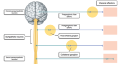
Label the general pattern of neurons and neurotransmitters associated with the autonomic nervous system.
The medulla oblongata is continuous caudally with the
Spinal cord
The primary function of the cerebellum is
Coordination of motor activity
Emotional responses are regulated in the
Hypothalamus
Respiration is regulated in the
Medulla oblongata
Gyri are characteristic of the
Cerebrum
Which meningeal layer follows the surface contours of the brain and spinal cord?
Pia mater
Separation of the periosteal and meningeal layers of dura mater forms
Dural venous sinuses
The subarachnoid space lies between the
Arachnoid mater and pia mater
The periosteal layer of dura mater is adherent to the
Inner surface of the skull
The subarachnoid space contains
Cerebrospinal fluid
Simple spinal reflexes occur independent of the brain.
True
Sensory stimuli enter the spinal cord via
Afferent axons
A simple spinal reflex typically involves how many neurons?
3
Interneurons are located in the
Spinal cord
Sensory receptors are found
Throughout the body
Aging of the brain begins
before birth.
Most cerebrospinal fluid is secreted from the choroid plexuses in the
lateral ventricles.
The simplest level of CNS function is the
spinal reflex.
A traumatic brain injury (TBI) results from
mechanical force.
The function of the cerebral association areas is
all of the above.
Melinda has Parkinson disease. Her movements are slowing and she has difficulty initiating voluntary muscular actions. The region that is affected in her brain is the
basal ganglia.
Basal ganglia are located in the ______ and ______.
deep regions of the cerebral hemispheres; aid in control of motor activities
Which of the following terms and definitions is correct?
Cerebral cortex-a thin layer of gray matter forming the outermost part of the cerebrum
Cerebrospinal fluid
all of the above.
The central nervous system (CNS) consists of
the brain and spinal cord.
The fourth ventricle is in the
brainstem
The part of the brain that coordinates voluntary muscular movements is the
cerebellum
Over the course of several months, Morris has experienced difficulty speaking coherently, clumsiness, muscle fasciculations, and increasing weakness in his limbs. These symptoms are most consistent with those of
amyotrophic lateral sclerosis.
Which lobe of your brain are you using when you answer this question?
Frontal
Spina bifida is a(n)
all of the above.
If the area of the cerebral hemisphere corresponding to Broca’s area is damaged,
motor control of the muscles associated with speech is lost.
Brain waves are recordings of activity in the
cerebral cortex.
The consequence of sensory nerve fibers crossing over is that the
right hemisphere of the cerebrum receives sensory impulses originating on the left side of the body and vice versa.
Aphasia is loss of the ability to
speak
Injury to the visual cortex of the right occipital lobe can cause
partial blindness in both eyes.
Which of the following are descending tracts in the spinal cord?
Rubrospinal
A lumbar puncture is
a test of the pressure that the cerebrospinal fluid is under.
Bob witnesses an auto accident and impulses from the ___________ division of the autonomic nervous system increase his heart rate.
sympathetic
Cerebrospinal fluid is produced by ______ and it __________.
choroid plexuses in the ventricles; protects the brain from blows to the skull
Remember! This essay was written by a student
You can get a custom paper by one of our expert writers
Order custom paper
Without paying upfront
