Merely a few decennaries into research, and the Zebrafish has unfolded an unbelievable sum of information that has helped non merely unravel the maps of legion cistrons, but has besides helped associate those cistrons to human upsets. The Zebrafish is a premier specimen to analyze. Their genome is to the full sequenced, 70 per centum of their familial codification lucifers worlds, and 84 per centum of human diseases have a Zebrafish parallels. New drugs and mutants are besides easy tested on Zebrafish since they absorb chemicals directly from their encompassing H2O. Zebrafish are besides easy handled in big Numberss, have a short coevals clip of three to four months, and can reproduce rapidly and in great figure. One of their more of import traits though, is that Zebrafish are semitransparent in their development phases so one can see organ constructions such as a beating bosom or go arounding blood. This is indispensable to cognize how a drug or mutant affects physiological traits.
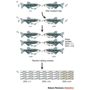 Forward and change by reversal genetic sciences are the footing for all familial screens done. Forward genetic science is used to place cistrons, or a set of cistrons, responsible for a certain phenotype of an being. Reverse genetic sciences is used to analyse the phenotype of an being following the break of a certain cistron. Research workers will exchange off cistrons, or present drugs to associate mutants to a alteration in phenotype or behaviour. In order to see how mutants affect the phenotype of Zebrafish, males must foremost be mutated and so mated to normal, wild type females ( F1 coevals ) . This creates the first coevals ( F2 coevals ) which is so crossbred to make the 2nd coevals ( F3 coevals ) . Some fish in the 2nd coevals will transport a homozygous mutant, holding two transcripts of the mutated cistron, one maternal and one paternal. Changes in phenotype can so be observed in these fish. These processs done with 1000s of mutated fish depict a big graduated table screen. The first big graduated table screen was done by two different groups in Boston and Tubingen. These groups found many cistron maps though this big graduated table screen including designation of written text factors necessary for endoderm formation such as Casanova ( Ca ) , bonnie and Clyde ( bon ) , and Faust ( fau ) . Besides, paired with the transparence of the Zebrafish, the groups were able to place cistrons that affect the cardiovascular system. The jekyll ( jek ) cistron was found to encode for the enzyme UDP ( uridine 5’- diphosphate ) – Glucose dehydrogenase which in bend is required for the formation of glycosaminoglycans. With glycosaminoglycans moving as the signaling tract jekyll forms the cardiac valve.
Other screens in Zebrafish such as the haploid and homozygous diploid screen helps place phenotypes due to recessionary allelomorphs in the F3 coevals without holding to test 1000s of fish. In this screen, wild type females are first fertilized by ethylnitrosourea mutagenized males. The ensuing female offspring are so fertilized with UV treated sperm making monoploid embryos. The UV radiation destroys the paternal DNA so the embryo merely carries the maternal DNA, and recessionary cistrons can be easier to place with ensuing progeny being 50 per centum mutation and 50 per centum wild-type.
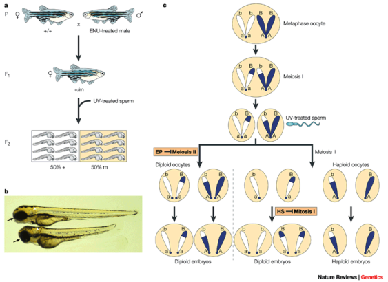
Another screen that is utilized is the mosaic screen used to increase the frequence of the mutant in involvement. In this screen pre-meiotic source cells from the grownup males are mutated through ethylnitrosourea mutagenesis. ENU merely alkylates bases on one Deoxyribonucleic acid strand, and the mutant merely becomes fixed during cell division after fertilisation. The resulting coevals is now Mosaic for a specific mutant, and when crossbred the resulting coevalss are mosaic for an estimated tenfold greater mutational burden. This rapid showing has helped place cistrons for cardiac development, and anterior and posterior patterning.
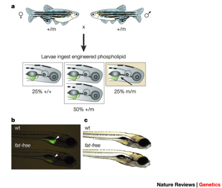
One really utile showing technique is to utilize fluorescent newsmans. This screen is particularly utile for mutant in specific enzymatic procedures. In the image below two fish heterozygous for a lipid lack, or fat free in the image, are bred and a one-fourth of their progeny will be homozygous for the recessionary digestive piece of land mutant. The fluorescent newsman is placed at the cleavage site at phospholipase A2. The ensuing tissue formation marked by the fluorescent newsmans show a decreased saddle sore vesica size, and physiological alterations in the digestive piece of land.
Though most screens are for physiological alterations, screens can besides place behavioural alterations. Zebrafish can be visually tested for their optokinetic response by puting a black and white membranophone in forepart of them, and so holding the membranophone rotate. Eye motions are recorded, and normal fish will scan the chevrons of the membranophone clockwise to counterclockwise, and so reset to the midplane before following the chevrons once more. Mutants in little or big eyes normally pass this trial, which suggests that oculus morphology has a different set of cistrons so ocular public presentation.
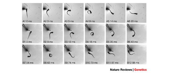
Normal larvae of three to four yearss will whirl and swim off from unwanted stimulations, but other than that are normally inactive. If the fish becomes a mutation for the cistron infinite plebe ( spc ) their flight behaviour from unwanted stimulations will alter, and the fish will turn on the topographic point, or even swim towards the unwanted stimulation. In the image below the times marked with A are the wild type Zebrafish, and the times marked with B are the infinite plebe mutations. Wild type fish will hold a C shaped spin, and so swim in the opposite way. Mutant fish will add another bend to their spin doing some to swim towards the unwanted stimulation.
Many human diseases have been better understood due to the research done on Zebrafish. Among the many mutants found in the Boston and Tubingen big screen, blood mutants were discovered that linked back to human upsets such as Erythropoietic Protoporphyria and Hepatoerythropoietic Porphyria. Erythropoietic Protoporphyria ( EPP ) is a blood upset that consequences in swelling, combustion, itchiness, and inflammation in the tegument after Sun exposure. The firing hurting normally ranges from mild to severe, and subsides in 12 to 24 hours. This disease normally manifests in early childhood, and is due to extra degrees protoporphyrin IX in red blood cells and plasma. The high degrees of protoporphyrin consequences from mutants in the ferrochelatase cistron ( fch ) , or in the delta- aminolevulinic acid synthase-2 cistron ( ALAS2 ) which is an X-linked mutant. Hepatoerythropoietic Porphyria ( HEP ) is more terrible than EPP and consequences in skin vesiculation. This type of porphyria is due to a lack in uroporphyrinogen decarboxylase ( UROD ) , and is an familial recessionary trait.


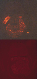
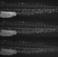 In the Boston Tubingen screen it was found that Dracula ( drc ) and yquem ( yqe ) mutations have red blood cell lysis when exposed to visible radiation. Figure 1 shows that after 120 seconds all red blood cells have been lysed. The yquem cistron encodes for uroporphyrinogen decarboxylase ( UROD ) , and homozygous mutations in this cistron have a lack in the UROD enzyme which leads to the more terrible HPP. Dracula encodes for ferrochelatase and when disrupted leads to erythropoietic protoporphyria in worlds. In another survey it was found that a homozygous drcm?48mutation has a transversion at a splice site of G bases to T in the ferrochelatase cistron. This creates an early halt codon, and produces the radiosensitivity phenotype. Other allelomorphs of the Dracula cistron were isolated including drcm87, drcm328, drcm248, and the m248 allelomorph showed liver abnormalcies which occurred even in dark raised embryos. Liver disease is non uncommon in patients with EPP since toxins are being absorbed from the lysed ruddy blood cells. Figure 2 shows the inclusions seeable in the liver of the drc mutation. Figure 3 shows that the inclusions are non limited to the vass, but are besides spread throughout the parenchyma. The mutant in these cistrons disrupts
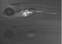 the haem biosynthetic pathway taking to defects in the last enzymes produced which leads to accretion of porphyrin intermediates, and it is these intermediates that when illuminated cause the production of free groups. This so leads to lipid peroxidation which affects the bilipid membrane of tissues including the cuticle. Due to the transparence and handiness, Zebrafish make for a perfect theoretical account for analyzing these porphyria diseases. Internal organs, such as the liver, can be monitored, and development can be watched to see how alterations in cistrons or add-on of drugs consequence red blood cell and corium development.
There is much more to detect in the field of genetic sciences, and scientists have merely begun to rub the surface with Zebrafish. With their easy handling, transparence in early development, short coevals clip, and parallels to human diseases, Zebrafish are premier specimen to analyze. Screening in Zebrafish such as a big graduated table screen, diploid homozygous haploid screens, mosaic screens, fluorescent newsman screens, and behavioural screens have helped in mapping out the Zebrafish’s genome and associating the maps of the cistrons to human procedures. Many human diseases have been better understood due to the survey of Zebrafish. Genes such as Dracula and Yquem have been identified in Zebrafish, and break of their cryptography for certain enzymes leads to blood upsets such as Erythropoietic Protoporphyria and Hepatoerythropoietic Porphyria. Through determination and understanding the maps of these cistrons research workers are better able to understand how EPP and HEP work, and how to forestall them.
Work Cited
Adams, Jill, and Kenna Shaw. “ Maping Genes to Chromosomes: Linkage and Genetic Screens. ”Nature.com. Nature Printing Group, 2008. Web. 05 Mar. 2014
& A ; lt ; hypertext transfer protocol: //www.nature.com/scitable/topicpage/mapping-genes-to-chromosomes-linkage-and-genetic-377 & A ; gt ; .
Childs, Sarah, Brant M. Weinstein, Manzoor-Ali Mohideen, Susan Donohue, Herbert Bonkovsky, and Mark C. Fishman. “ Zebrafish Dracula Encodes Ferrochelatase and Its Mutantion Provides a Model for Erythropoietic Protoporphyria. ”Current Biology10.16 ( 2000 ) : 1001-004. Web. 5 Mar. 2014. & A ; lt ; hypertext transfer protocol: //www.sciencedirect.com/science/article/pii/S0960982200006539 & A ; gt ; .
“ Erythropoietic Protoporphyria ( EPP ) or Protoporphyria. ”American Porphyria Foundation. American Porphyria Foundation, 2010. Web. 05 Mar. 2014.
& A ; lt ; hypertext transfer protocol: //www.porphyriafoundation.com/about-porphyria/types-of-porphyria/EPP & A ; gt ; .
“ Hepatoerythropoietic Porphyria ( HEP ) . ”American Porphyria Foundation. American Porphyria Foundation, 2010. Web. 05 Mar. 2014.
& A ; lt ; hypertext transfer protocol: //www.porphyriafoundation.com/about-porphyria/types-of-porphyria/HEP & A ; gt ; .
McKie, Robin. “ How the Diminutive Zebrafish Is Having a Big Impact on Medical Research. ”The Observer. Guardian News and Media, 15 Sept. 2013. Web. 05 Mar. 2014.
& A ; lt ; hypertext transfer protocol: //www.theguardian.com/science/2013/sep/15/zebrafish-human-genes-project & A ; gt ; .
Patton, Elizabeth E. , and Leonard I. Zon. “ The Art and Design of Genetic Screens: Zebrafish. ” ( n.d. ) : n. pag. Web. 3 Mar. 2014. & A ; lt ; hypertext transfer protocol: //www.rz.uni-karlsruhe.de/~db45/Studiendekanat/Lehre/Bachelor/Modul_06A/Material/Zebrafish.pdf & A ; gt ; .
Pearson, Helen. “ Submerged Space Cadet. ”Nature.com. Nature Printing Group, 01 June 2001. Web. 05 Mar. 2014.


