- . Introduction
1.1. Cancer
Cancer is one of the prima causes of mortality and is the causal factor of every bit many as 25 % of deceases in the United States ( ACS, 2014 ) . In general, malignant neoplastic disease is a group of different diseases characterized by aberrant, unregulated cell growing. In malignant neoplastic disease, due to loss of control of cell division and growing, formation of balls or tissue multitudes, termed tumours, occurs. The malignant neoplastic disease may besides migrate to other parts of the organic structure via the blood or lymphatic systems, taking to devastation of healthy tissues. The formation of blood cells basically takes topographic point in the bone marrow and is a closely regulated procedure affecting proliferation, distinction and cell endurance. Cancers arise from a gradual buildup of familial alterations due to clonal enlargement events in pre-malignant cell populations that are undergoing Darwinian choice procedure ( Weinberg, 2007 ) .
1.2. Hematopoiesis
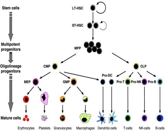
Hematopoiesis is a important procedure in the growing and re-generation of blood cells, since these cells are indispensable for the ordinance of assorted procedures such as immune response, supply of O to tissues and homeostasis. The haematopoietic system comprises of blood cells, bone marrow, lymph node, Thymus, spleen and endothelial cells. The Precursor Hematopoietic root cell ( HSC ) is a pluripotent root cell, capable of spliting and giving rise to cells of different line of descents. On having distinction stimulation, HSC divides to give rise to Multipotent Progenitor cell ( MPP ) , which does non hold self-renewal capacity. This cell divides to give rise to precursor cells of lymphoid and myeloid line of descents ( Figure 1.1 ) . The common lymphoid precursor cell in bend gives rise to Pro-B, Pro-T and Pro-NK cells which divide to organize B- , T- and NK-cells. The common myeloid precuror cell gives rise to the megakaryotic/erythroid primogenitor which farther divides to organize red blood cells and thrombocytes, Myelomonocytic primogenitors which divide to organize granulocytes and macrophages. Both the common lymphoid precursor and the common myeloid precursor cell may besides give rise to Pro-dendritic cell which divides to organize dendritic cell ( Passegue et al. , 2003 ) . Carcinogenic stimulation may take to changes in this procedure, transforming a normal haematopoietic root cell precursor to a leukemic root cell. Therefore, deviant haematopoiesis leads to development of leukaemia.

Figure 1.1. Conventional overview of haematopoiesis[ LT-HSC= Long Term Hematopoiestic Stem Cell, ST-HSC= Short Term Hematopoietic Stem Cell, MPP= Multipotent Progenitor, CMP= Common Myeloid Progenitor, CLP= Common Lymphoid Progenitor, MEP= Megakaryotic/Erythroid Progenitor, GMP = Myelomonocytic Progenitor, Pro-DC = Dendritic Cell Progenitor, Pro-T = T-cell Progenitor, Pro-NK= Natural Killer cell Progenitor, Pro-B= B-cell Progenitor ] ( Source: Passegue et al. , 2003 )
1.3. Leukemia
Cancer has been known to impact many organic structure variety meats and tissues, some of them being castanetss, tummy, lungs and blood. The malignant neoplastic disease of the blood is called Leukemia. It is a subtype of a wide array of diseases normally referenced as Hematological Malignancies. Harmonizing to statistics provided by the Leukemia and Lymphoma Society, this disease is expected to be diagnosed in more than 52,380 people in the United States in 2014, and it has one of the top mortality rates among different types of malignant neoplastic disease ( Leukemia & A ; Lymphoma Society, 2014 ).Based on the badness of the disease and the type of white blood cell affected, leukaemia is classified into different types.
1.4. Types of leukaemia
Most leukaemias may be subdivided into two general groups: myeloid leukaemia ( ~60 % of the instances ) and lymphocytic leukaemia ( ~40 % of the instances ) . Leukemias are besides classified based on whether they are acute ( ~55 % ) or chronic ( ~45 % ) . In acute leukaemia, the malignant cells, or blasts, are immature cells that have lost their ability to distinguish and are therefore incapable of executing their maps in immune response. The oncoming of acute leukaemia is rapid, by and large hebdomads, and, is largely fatal, unless the intervention is initiated fleetly. Chronic leukaemia occur in mature cells and consequence in decrease in their operation capacity. These unnatural cells besides proliferate at a slower rate, by and large old ages. Therefore, leukaemias may be categorized into four chief types: Acute Lymphocytic Leukemia ( ALL ) , Chronic Lymphocytic Leukemia ( CLL ) , Acute Myelogenous Leukemia ( AML ) , Chronic Myelogenous Leukemia ( CML ) , each of which farther comprises several subtypes. Our survey is focused on ALL.
1.5. Acute Lymphocytic Leukemia ( ALL )
Acute lymphoblastic ( lymphocytic ) leukaemia ( ALL ) comprises of a group of lymphoid tumors that have indistinguishable morphological and immunophentoypical features to precursor cells of B- and T-lineages. These tumors may show mostly as a complete leukemic procedure, with widespread engagement of the bone marrow and peripheral blood cells or they may be restricted to weave incursion, with really small ( & lt ; 25 % ) to no bone marrow engagement. The former instance is classically designated as lymphoblastic lymphomas ( LBLs ) . ALL and LBLs appear to consist a biologic gamut, although they may show distinguishable clinical characteristics. The current World Health Organization ( WHO ) Classification of haematopoietic malignant neoplastic diseases delegates these upsets as B- or T-lymphoblastic leukemia/lymphoma ( Swerdlow et al. , 2008) .
1.6. Clinical symptoms and diagnosing of ALL
The chief symptoms of ALL include: febrility, anemia, increased hemorrhage and bruising, shortness of breath, infections and bone and joint hurting. Initial diagnosing of ALL includes appraisal of complete blood image with analysis of entire count of ruddy blood cell, white blood cell and thrombocyte Numberss, as these Numberss are by and large altered in ALL ( Daly et al. , 2010 ) . By and large, Children with ALL have low thrombocyte count with low ruddy blood cell ( hemoglobin ) degrees and concomitantly high white cell count, with a excess of immature blast cells. These blast cells do non distinguish and maturate ; alternatively they proliferate in inordinate Numberss and besides prevent development and operation of normal unchanged blood cells. ALL is confirmed via appraisal of the per centum of blast cells in the patient’s.bone marrow. Under normal physiological conditions, there are less than 5 % blast cells present in healthy persons. In ALL patients, blast cell scope between 20 % – 95 % is by and large reported ( Daly et al. , 2010 ) . Of the two types of white blood cells affected by ALL, B-cell ALL constitutes about ~85 % and T-cell ALL constitutes ~15 % of ALL instances. Both of these subtypes are diagnosed by measuring the morphology of cells in blood or bone marrow specimens collected from the patients ( Daly et al. , 2010 ) . ALL has been differentiated into subtypes based on morphology, cytochemistry and immunophenotyping. The conventional standards used to sort ALL are based on categorization system of the French-American-British ( FAB ) group ( Bennet et al. , 1976 ) . The FAB group defines the 3 subtypes of ALL ( L1, L2 and L3 ) based on the morphological characteristics of the blasts when viewed under a microscope. This categorization is based on ratio of nucleole to cytoplasm, presence and size of nucleole, grade of consistence in the form of the atomic membrane and size of the cell ( Bennett et al. , 1981 ) . The ALL-L1 subtype comprises chiefly little size blast cells and is found in 70-80 % of childhood ALL instances. ALL-L2 subtype comprises of a assorted group of little and big blast cells, with a higher per centum of big sized cells. The ALL-L3 subtype comprises of medium to big sized blast cells. In contrast to this categorization system, The European Group for the Immunological Classification of Leukemias ( EGIL ) classifies acute leukaemias entirely on the footing of immunophenotyping ( Abdul-Hamid, 2011 ) .
1.7. Incidences of Acute Lymphoblastic Leukemia ( ALL )
Harmonizing to WHO Report in 2003, 250,000 instances of leukaemia were reported. En masse, leukemias history for approximately 31 % of all childhood malignant neoplastic diseases and impact approximately 2,200 American kids and immature grownups each twelvemonth and consequences in decease in 3.0 % ( 618 under the age of 19 ) of these instances. In England and Wales about 400 kids are diagnosed each twelvemonth and about 100 dices due to leukemia ( Shah and Coleman, 2007 ) . The mean incidence of leukaemia in kids in the European Region was 46.7 instances per million per twelvemonth ( WHO, 2009 ) . ALL is the most normally diagnosed malignant neoplastic disease in kids, accounting for 26 % of malignant neoplastic diseases diagnosed in those elderly birth to 14 old ages. ALL is more common in industrialised states than in developing states. The incidence rate of childhood ALL in USA is approximately 35 to 40 instances per million ( Howlader et al. , 2013 ) . The incidence rates of a few ethnicities, metropoliss and states have been represented in Table 1.1 ( Ross et al. , 2011 ) . In India, approximately 25 people per million population are affected yearly ( about 500 instances per twelvemonth ) with comparative proportion of ALL changing between 60 and 85 % of all leukaemia found in kids. The mortality rate is reported to be high with merely 33 % lasting at five old ages ( Bombay Cancer Registry 1996, Arora et al. , 2009 ) . Arora et Al. ( 2009 ) , through their meta-analysis, reported a higher incidence of T-ALL ( 20-50 % ) in India when compared to the developed states, along with the more frequent presence of hypodiploidy and T ( 1 ; 19 ) , t ( 9 ; 22 ) , and T ( 4 ; 11 ) translocations in Indian childhood ALL. Besides, based on their analysis of the malignant neoplastic disease register, they observed a somewhat higher incidence of ALL in male kids than in female kids.
Table 1.1. Incidence rates of Acute Lymphoblastic Leukemia ( ALL ) ( Ross et al. , 2011 )
|
State |
Incidence rate/million |
|
US Spanish americans |
49.9 |
|
Costa Rica |
46.3 |
|
US White persons |
45.4 |
|
Greece |
44.9 |
|
Mexico |
44.5 |
|
The Netherlands |
30.9 |
|
Lima/Peru |
25.4 |
|
US Blacks |
18.7 |
|
Bombay/India |
16 |
|
Uganda/Africa |
3.3 |
1.8. Familial Aspects of Acute Lymphoblastic Leukemia ( ALL )
Present diagnostic methods reveal that familial aberrances occur in about 90 % of ALL patients. In most instances these aberrances were found to be specific to the leukaemia type and besides to immunological or morphological leukaemia subtypes. In ALL, many of the familial disturbances are clearly different in kids and grownups ( Ma et al. , 1999 ) . Research on ALL, utilizing familial, proteomic, look and genome broad association surveies, have shown that the normal biological procedures and cell development and distinction tracts such as cell rhythm, proliferation, cellular signaling, haematopoiesis, epigenetic ordinance are deregulated in leukemic cells due to changes in the cistrons and proteins involved in these procedures ( Pui et al. , 2012 ) . Focal omissions and mutants in the written text factorsPAX5,IKZF1( Mullighan et al. , 2007 ) , written text regulatorCREBBP( Mullighan et al. , 2011 ) , protein tyrosine kinasesJAK1,JAK2( Mullighan et al. , 2009 ) , cell rhythm regulator and tumour suppresserTP53( Hof et al. , 2011 ) , chromosomal rearrangements inCRLF2( involved in haematopoiesis ) ( Mullighan et al. , 2011 ) have been observed in leukemic cells. Further, changes in cell rhythm regulators such as cyclin D1 ( Aref et al. , 2006 ) and haematopoietic regulators such as Notch1 ( often mutated in T-ALL, Weng et al. , 2004 ) have besides been reported. Zhang et al. , ( 2011 ) besides reported mutants in RAS signaling, JAK/STAT and B-cell development tract. Surveies have besides shown that changes inFLT3cistron play an of import function in leukemogenesis ( Reddy et al. , 2006a ) . Single nucleotide polymorphisms ( SNPs ) in cistrons such as vitamin Bc metabolizingMTHFR( Reddy and Jamil, 2006b ) ,RFC1,NNMT( de Jonge et al. , 2009 ) , xenobiotic metabolizingCYP1A1*2A,GSTM1void type ( Krajinovic et al. , 1999 ; Reddy and Jamil 2006c ) ,NAT2( Krajinovic et al. , 2000 ) ,CYP2E1,MPO, NQO1( Krajinovic et al. , 2002 ) ,GSTT1, immune map cistronsIL12A( Chang et al. , 2010 ) ,HLA-DPB1*0201( Taylor et al. , 2002 ) were found to be associated with increased hazard of developing ALL, particularly in kids. These surveies emphasize the critical function played by changes in cistrons and proteins in neoplastic transmutation of ALL and therefore point to the demand to better understand the biomolecules involved in the ordinance of important deregulated tracts such as cell rhythm and haematopoiesis that are normally aberrated in ALL.
1.9. Cytogenetic changes of Acute Lymphoblastic Leukemia ( ALL )
Cytogenetic aberrances are presently one of the major predictive factors in ALL. Cytogenetic surveies have reported legion chromosomal aberrances in patients with ALL. These include changes in chromosome figure ensuing in High Hyperdiploidy with 51 to 65 chromosomes per cell and Hypodiploidy with less than 44 chromosomes. Changes in chromosomal construction, chiefly translocations, have besides been observed, including ETV6-RUNX1 ( T ( 12 ; 21 ) , Philadelphia chromosome ( T ( 9 ; 22 ) , MLL translocations and TCF3-PBX1 ( besides known as E2A-PBX1 ; T ( 1 ; 19 ) ) . These chromosomal figure and construction changes, together with the other predictive factors, have been observed to impact intervention response.
1.10. Rationale for Bioinformatics attack
In recent old ages, experimental research has been supplemented to a big extent with the usage of computational attacks. The application of bioinformatics methodological analysiss has helped in deriving new penetrations into disease biological science, particularly in malignant neoplastic diseases, through feasibleness of big graduated table informations analysis in lesser clip. Besides, in silico methodological analysiss offer a huge array of informations analysis tools that make it executable to analyze biological informations via application of multi-parameter testing. Phylogenetics package have helped in understanding evolutionary forms of cistrons among the different life beings. These forms may keep the hint to decode the changes of cistrons in human disease, through a comparative survey of similar cistrons in other beings as demonstrated in our surveies ( Jayaraman et al. , 2011 ; Jayaraman and Jamil, 2012 ) . Further, computational methods are particularly utile in analysis of microarray look informations. Data generated from look surveies are by and large voluminous and their illation manually would necessitate a batch of clip and resources and may besides be capable to manual mistakes. The usage of computational algorithms ensures the handiness of multiple analysis parametric quantities that search the information for forms rapidly and exhaustively and therefore aid in bring forthing extended consequences that can be interpreted more accurately. Besides, application of cistron prioritization algorithms have become highly utile in shortlisting new cistron marks in disease from an extended set of plausible campaigners, therefore contracting down the hunt for placing new curative marks in disease research. Further, in many complex diseases, particularly malignant neoplastic diseases, cistrons do non work in isolation, but are portion of immense interconnected web tracts that act as a disease main road and lead to a cascade of pathogenetic alterations. Understanding the participants in these webs through bioinformatics tools is a more executable attack since mapping each of these connexions by experimentation would merely be possible through the usage of thorough resources and extended clip period. Computational anticipation of networking integrates multiple information beginnings such as available experimental informations, informations from related species and happening upstream or downstream of a peculiar tract and similar maps to map cistrons and their proteins to web faculties. We have utilizedin silicoprotein networking to clarify interactors of TP53 and NOTCH1 and their function in leukemogenesis ( Jamil et al. , 2012 ; Jayaraman and Jamil, 2013 ) .
Pharmacogenomics is a field that correlates genetic/genomic information of persons to pharmacological information to find the best curative regimen for a peculiar individual. This seamster made therapy is highly indispensable since non all persons respond the same manner to intervention. Pharmacogenomics has been highly utile in Oncology in designation of drugs and finding of drug efficaciousnesss, e.g. in designation of efficaciousness of the drug Trastuzumab in HER2 positive chest malignant neoplastic disease with metastasis ( Shak, 1999 ) , non-efficacy of the drug 6-Mercaptopurine in leukaemia and lymphoma patients with certain polymorphisms in TPMT enzyme, designation of inefficaciousness of Cetuximab, aiming EGFR, in patients with KRAS-induced malignant neoplastic diseases ( Weng et al. , 2013 ) . The application of bioinformatics techniques in pharmacogenomics has been highly utile as computational attacks have furthered our apprehension of the genes/proteins involved in disease and besides aided designation of new drugs. Homology patterning and in silico molecular docking utilizing computational package have provided the agencies to visualise protein and Deoxyribonucleic acid construction in 3-dimensional infinite, survey whether a peculiar drug may adhere to the protein or Deoxyribonucleic acid and if so, look into how effectual the binding is and visualise the interactions of the drug with the aminic acids of the protein and the bases of the DNA. Computational attacks have become indispensable in drug find research, since traditional drug find methods normally take a really long clip from designation of new drug to let go of in the market. Application of bioinformatics attacks to drug find have helped rush up release of effectual drugs in the market and have besides helped in cost decrease by important degrees ( Song et al. , 2009 ) . Further, they have been vastly utile in foretelling possible toxicity of drugs, therefore assisting in development of more powerful but less toxic drugs. Besides, the application of bioinformatics methods as an initial measure in disease research would assist in contracting down the research inquiries prior to experimental work and therefore aid in hastening research surveies and salvaging resources. Therefore, the usage of bioinformatics tools and techniques has been proved to be really utile in fostering our apprehension of disease biological science and hence has been applied in our survey to deduce information about leukemogenesis in ALL. Hence, the aim of this survey was to analyse the cistrons and tracts that are known to be deregulated in ALL utilizing bioinformatics tools to deduce new information with respect to their function in leukemogenesis, to profile new cistrons based on interconnectivity with already bing leukaemia associated cistrons and research their usage as possible markers of predictive and diagnostic involvement and to execute in silico drug adhering surveies to deduce new marks for therapy.


