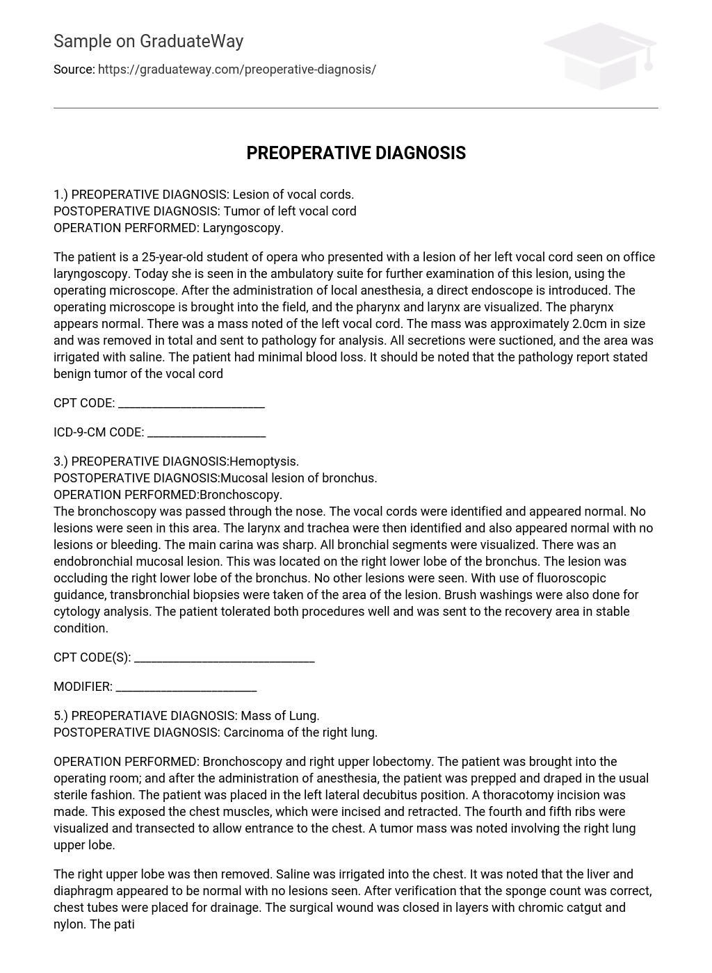
PREOPERATIVE DIAGNOSIS: Vocal cord lesion.
POSTOPERATIVE DIAGNOSIS: Tumor of left vocal cord
OPERATION PERFORMED: Laryngoscopy.
A 25-year-old opera student went to the office for a laryngoscopy and a lesion was found on her left vocal cord. Today, she is at the ambulatory suite to evaluate the lesion further using an operating microscope. Local anesthesia is given before inserting a direct endoscope.
Using the operating microscope, both the pharynx and larynx are visualized. While the pharynx appears normal, a mass is discovered on the left vocal cord measuring around 2.0cm. The mass is successfully removed for further analysis. Secretions are eliminated with suction and the area is irrigated using saline solution. Remarkably, there is minimal blood loss during the procedure. Subsequently, pathology examination confirms that the tumor on the vocal cord is benign.
Preoperative Diagnosis: The patient is experiencing hemoptysis.
POSTOPERATIVE DIAGNOSIS: The patient had a bronchial mucosal lesion.
PROCEDURE: Bronchoscopy was carried out.
Throughout the bronchoscopy procedure, the device was introduced via the nasal passage to examine different sections of the respiratory system. The vocal cords, larynx, and trachea displayed no abnormalities or lesions. The main carina, where the trachea divides into the bronchi, appeared to be sharp. All segments of the bronchial tree were observed without any lesions or bleeding. Nonetheless, a lesion was discovered in the inner mucosal lining of the right lower lobe bronchus.
The bronchus in the right lower lobe was obstructed by the lesion, while no other lesions were observed. Under the guidance of fluoroscopy, biopsies were conducted on the affected area through the bronchial wall. Additionally, brush washings were performed to obtain samples for cytology analysis. The patient underwent both procedures successfully and was subsequently transferred to the recovery area in a stable condition.
PREOPERATIVE DIAGNOSIS: Mass of Lung.
POSTOPERATIVE DIAGNOSIS: Carcinoma of the right lung.
OPERATION PERFORMED: Bronchoscopy and right upper lobectomy.
The patient was brought into the operating room and administered anesthesia. They were then prepped and draped in the customary sterile manner. Placed in the left lateral decubitus position, a thoracotomy incision was made, exposing the chest muscles. The incised and retracted muscles revealed the fourth and fifth ribs, which were transected to gain access to the chest. Notably, a tumor mass was found affecting the upper lobe of the right lung.
The surgical procedure involved performing a right upper lobe removal and rinsing the chest with saline solution. The liver and diaphragm were found to have no abnormalities or lesions. After confirming the correct number of sponges, drainage chest tubes were inserted and the surgical wound was closed using chromic catgut and nylon stitches. No complications occurred during this stage while the patient was under anesthesia.
Following that, while still under anesthesia, the patient’s position was adjusted to their back for a bronchoscopy. A flexible fiberoptic bronchoscope was used to examine clear airways on both sides. Once the scope was withdrawn, the patient regained consciousness before being transferred to a stable recovery area.
Preoperative Diagnosis: Acute respiratory insufficiency caused by ALS.
POSTOPERATIVE DIAGNOSIS: Acute respiratory insufficiency due to ALS
OPERATION PERFORMED: Tracheostomy.
A 45-year-old male with ALS is suffering from severe progressive shortness of breath. After assessing the potential risks, it was decided that a tracheostomy would be conducted on him. The patient was brought to the operating suite and positioned supine on the table for the procedure. General anesthesia was given, and the patient was prepared and covered using standard sterile procedures.
In the neck, a 2.5cm cut was made over the trachea. The trachea was carefully separated from the surrounding structures once the tracheal rings were located. After finding the second ring, a tube was inserted through the incision. The patient’s breath sounds were assessed and found to be sufficient. Gauze was used to pack the tracheostoma and the ties were secured. After the operation, a chest x-ray will be performed to confirm proper tube placement; however, the patient had good breath sounds upon transfer to the recovery room.
LOCATION: Outpatient. Hospital
PATIENT: Liz Charles
PHYSICIAN: Gregory Dawson. MD
STUDY PERFORMED BY PHYSICIAN ONLY:Nocturnal polysomnogram without CPAP titration
ENTRANCE DIAGNOSIS: Somnolence
This is a fully attended, multichannel nocturnal polysomnogram, with the patient spending 386.6 minutes in bed and 317 minutes asleep. They experienced 61 arousals throughout the night, which is higher than normal and suggests difficulty with sleep maintenance. The patient took 18.5 minutes to fall asleep and had a REM latency of 171.5 minutes, both slightly prolonged. They experienced 27 respiratory events during the night, including obstructive apneas and obstructive hypopneas, resulting in a respiratory disturbance index of 5.1. A respiratory disturbance index over 5 is considered significant. The longest duration of any individual event was 34 seconds. The patient’s O2 saturation levels ranged from 76% to 95%, with 29% of the time spent with O2 sats below 88%.
During observed obstructive events, the patient’s heart rate fluctuated between 55 and 113, demonstrating minimal changes. Grade 1-2 snoring was documented, with respiratory disturbance events being most noticeable during REM sleep while in a supine position. All five stages of sleep were identified. The sole abnormality discovered was a reduction in the duration of REM sleep.
OVERALL IMPRESSION: The patient exhibited a significant level of obstructive sleep apnea, as evidenced by a respiratory disturbance index of 5.1 indicating substantial disruption. Moreover, it was observed that the patient spent 29% of the time with oxygen levels below 88%, further confirming the existence of obstructive sleep apnea. It is recommended to arrange a follow-up session for CPAP titration in order to address this condition. To sum up, the overall impression remains consistent with obstructive sleep apnea.


