


Quantifying protein concentration is a really important procedure for analysing protein. It is indispensable in order to place, qualify, and sublimate proteins, and this can besides be usage in medical researches by helping in diagnosing of certain diseases. There are several of methods that can be usage for protein quantification. Each has its ain advantages and disadvantages. One of which is the Bradford check, which is normally used because of its simpleness, broad estimated working scope, and sensitiveness to molecules of involvement.
Chicken egg white albumen was used as a protein beginning. Different concentrations of albumen were prepared and their several optical densities at 595nm were obtained. This assay uses Coomassie Blue G-250 dye that forms a complex with certain aminic acids. A standard curve was created from the natural information of optical density reading. From the Pearson’s Rho correlativity, it was shown that the optical density and concentration has a strong positive relationship. The chemical reactions involve in this check was besides analyzed and understood.
Introduction
Proteins are really of import in an being for growing and saving. Before analysing a protein, it requires to find the measure of proteins present. The consequence of this will be utile in word picture and purification of proteins, in designation and in diagnosing of diseases, since some unwellnesss affect the degree of proteins. There are different methods in order to quantify protein concentration. It is of import for an check to be able to be applied to a broad scope of concentrations. It would besides be good for the check to be sensitive plenty to observe even the smallest protein content in order to hold an accurate consequence.
The check should besides be specific to the constituent that is to be quantified. This is to avoid contaminations to be detected, such as cell constituents, supermolecules like saccharides, nucleic acid and lipoids. There are different methods on quantifying proteins. One of which is the Non-colorimetric processs. This includes finding of N derived from proteins, analysis of amino acids, and happen out of dry affair stuff of protein. The 1 that is common in this modern clip is the Colorimetric methods of quantitation. This is because of the technological promotions available like use of spectophotometers ( Ninfa et al. , 2009 )
In biuret check, cupric ( Cu2+ ) ions are being reduced to cupric ( Cu1+ ) ions by the proteins. This cupric ions forms a complex with the peptide bonds giving a bluish colored complex. This check requires high concentration of proteins since it is rather insensitive ( Dennison, 2003 ) .
Lowry check starts with a protein-copper complex merely like the merchandise in the Biuret check. In Lowry assay it is so followed by the decrease of Folin–Ciocalteu reagent under alakaline conditions. Cupric ions are the 1s involve in the procedure of decrease, ensuing to a intense blue colour. Lowry check is more sensitive unlike the biuret check ; nevertheless, since it’s really sensitive, it can observe other constituents that are non of involvement merely like detergents ( Dennison, 2003 ) .
BCA Assay is the same as the Lowry check, but bichoninic acid ( BCA ) is the 1 being reduced by the protein alternatively of Folin–Ciocalteu reagent. BCA check is sensitive but non to other contaminations. It is more sensitive to saccharides, lipoids and other substances ( Dennison, 2003 ) .
Bradford check, which is the most normally used colorimetric method, uses Coomassie Blue G-250 dye. This dye forms a noncovalent bond with proteins chiefly basic amino acids ( arginine, lysine and histidine ) . This complex consequences to a bluish signifier in colour. This check is sensitive, accurate and can be done rapidly ( Redmile-Gordon et al. , 2012 ) .
The beginning of protein for this experiment is the poulet egg white albumen. Studies dwelling of iron-chelation, peptidase suppression, immunoregulation, etc. uses lily-livered egg white albumen every bit good. Purification of the albumen is really much needed before making any experimental process with the protein ( Geng et al. , 2012 ) . In fact the albumen is dwelling of 385 amino acid residue ( Alleoni, 2006 ) .
In finding the protein concentration, it is really of import to do a standard curve in every check perfumed. Pearson’s Rho correlativity is used to verify the additive relationship between the two variables involve in this experiment, optical density and albumin concentration ( Statstutor, n.d. ) .
This survey aims to verify how the concentration of proteins can impact the optical density of the sample. It is besides to do a standard curve for Chicken egg albumen and to cognize the Pearson’s Rho Correlation. It is besides really of import to understand the procedures involved with protein quantification. This experiment besides makes one to be excess careful with the research lab processs so that accurate informations can be obtained. In a higher concentration, there are more atoms involved, and so when a UV visible radiation work stoppages, more atoms will be able to absorb it and higher optical density value will be obtained.
MATERIALS AND METHODS
Before anything else, the UV/VIS Spectrophotometer was turned on before get downing the processs in order for the machine to warm up and map decently this avoids interfering with the informations reading. Adequate sums of Chicken egg white albumen 10mg/mL, Phosphate Buffered Saline ( PBS ) pH 7.4, and Bradford reagent were obtained in a beaker from the reagent bottle.
The beaker which contained the Bradford reagent was covered with paper since the the reagent is really light sensitive and might impact the informations. With the usage of micropipettors, albumen, phosphate buffered solution and Bradford reagent with known concentrations were transferred to six different microfuge tubings severally. The concentrations of the reagents were the followers:
Table 1: Volume of stock solutions for the readying of different albumen concentrations
| Standard no. | Bradford Reagent ( ul ) | Phosphate Buffered Saline ( ul ) | Chicken Egg White Albumin ( ul ) | Concentration ( ug/ul ) |
| Space | 500 | 500 | 0 | |
| 1 | 500 | 420 | 80 | 0.8 |
| 2 | 500 | 340 | 160 | 1.6 |
| 3 | 500 | 260 | 240 | 2.4 |
| 4 | 500 | 180 | 320 | 3.2 |
| 5 | 500 | 100 | 400 | 4 |
The solutions were vortex one at a clip for 10 seconds. It settled for 10 proceedingss. After that, the solutions were once more vortex for 5 seconds. Then the solutions from the microfuge tubings were transferred to the cuvettes separately. The cuvette was non touched on the level side panel. The cuvettes were gently placed in the spectreophotometer. The optical density of each concentration was read at 595 nanometer. The processs were done for two tests.
The standard curve and finding of Pearson’s Rho correlativity were made from the natural information of optical density readings.
Consequence
Table 2: Series of known concentrations of Albumin
| Standard no. | Bradford Reagent ( ul ) | Phosphate Buffered Saline ( ul ) | Chicken Egg White Albumin ( ul ) | Concentration ( ug/ul ) |
| Space | 500 | 500 | 0 | |
| 1 | 500 | 420 | 80 | 0.8 |
| 2 | 500 | 340 | 160 | 1.6 |
| 3 | 500 | 260 | 240 | 2.4 |
| 4 | 500 | 180 | 320 | 3.2 |
| 5 | 500 | 100 | 400 | 4 |
From the volume of the stock solutions, different concentrations of albumen were made. The albumin concentration of the criterions ranges from 0.0- 4.0 ug/ul and have an increase of 0.8.
Table 2: Pearson’s Rho Correlation of the Absorbance readings at 595nm and Albumin Concentrations
- Trial 1 ; ( B ) Trial 2 ; ( degree Celsius ) Mean optical density of the two tests
A.
| Standard no. | Concentration ( ug/ul ) | Trial 1 ( A ) |
| Space | 0.0000 | 0.0000 |
| 1 | 0.8000 | 0.7170 |
| 2 | 1.6000 | 0.6750 |
| 3 | 2.4000 | 0.3790 |
| 4 | 3.2000 | 0.7190 |
| 5 | 4.0000 | 0.8640 |
| Pearson ‘s R Correlation | 0.679886127 | |
B.
| Standard no. | Concentration ( ug/ul ) | Trial 2 ( A ) |
| Space | 0.0000 | 0.0000 |
| 1 | 0.8000 | 0.0510 |
| 2 | 1.6000 | 0.5480 |
| 3 | 2.4000 | 0.7740 |
| 4 | 3.2000 | 0.7990 |
| 5 | 4.0000 | 0.4250 |
| Pearson ‘s R Correlation | 0.710839544 |
C.
| Standard no. | Concentration ( ug/ul ) | Average optical density ( A ) |
| Space | 0.0000 | 0.00 |
| 1 | 0.8000 | 0.3840 |
| 2 | 1.6000 | 0.6115 |
| 3 | 2.4000 | 0.5765 |
| 4 | 3.2000 | 0.7590 |
| 5 | 4.0000 | 0.6445 |
| Pearson ‘s R Correlation | 0.84783844 |
The optical density value of the five albumens concentrations were measured twice. With respects to the Pearson’s Rho Correlation of each, Trial 1 shows that the Albumin concentration and Absorbance at 595nm has a strong positive relationship. While test 2 and the mean of the two tests show that the two variables exhibit a really strong positive relationship.
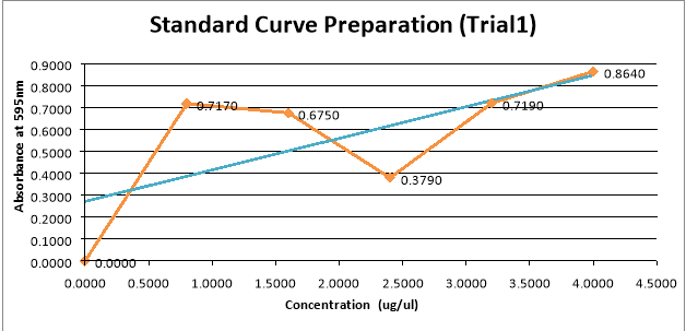
Figure 1: Trial 1 Optical density at 595nm V. Concentration of Albumin ( ug/ul )
In test 1, the Peason’s Rho Correlation value is 0.679886127 demoing a strong positive relationship between the two variables. From the optical density value of the standard no.1, there is a lessening in the optical density in standard no.2 and criterion. No.3. Then a sudden addition in optical density in standard no.4 and standard no.5. And besides from the line equation, the incline has a positive value giving an upward way and a direct correlativity between optical density and concentration of the albumen.
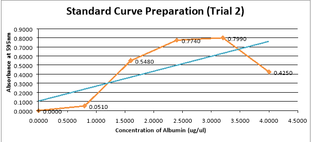
Figure 2: Trial 2 Optical density at 595nm V. Concentration of Albumin ( ug/ul )
Trial 2 shows a really strong positive correlativity between the two variables since the value of the Pearson’s Rho Correlation value is 0.710839544. Standard no.1 until Standard no.4 shows that the optical density at 595nm is increasing with the albumin concentration. But for standard no.5, the optical density of 4.00 ug/ul Albumin went down really rapidly. Based on the line equation of the best fit line of this graph, the incline has a positive value giving an upward way and a direct correlativity between optical density and concentration of the albumen.
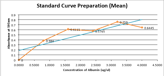
Figure 3: Average Absorbance of the two tests at 595nm V. Concentration of Albumin ( ug/ul )
The average values of the optical density at 595nm for the two tests were obtained and graph. The average optical density and albumen concentration shows a strong positive correlativity, holding a Pearson’s Rho correlativity of 0.84783844. The graph shows that there are two outliers, standard no. 3 and standard no.4. A direct correlativity between average optical density and concentration of the albumen is besides shown based on the incline of the line equation.
Discussion
Bradford assay utilize Coomassie Blue G-250 dye that forms a complex with the basic amino acids and therefore holding a bluish signifier in colour as the result ( Redmile-Gordon et al. , 2012 ) .
The concentration of the poulet egg white albumen ranges from 0.0 ug/ul, which is the space, up to 4.0 ug/ul. This shows an increasing concentration of the albumen, and to hold an accurate informations, there is 0.8 so that the spread between the concentrations is equal. With this, the optical density at 595nm can be compared to see the relationship between the two variables.
In Pearson’s Rho Correlation, the closer the value to 1 or -1, the strong is the additive correlativity for the two variables ( Statstutor, n.d ) . In test 1, the Pearson’s Rho Correlation value is 0.679886127, and since the value ranges from +.40 to +.69, the Albumin concentration and optical density value has a strong positive. The graph shows that the optical density readings are non consistent. Standard no.2 and criterion no.3 shows a sudden lessening.
The Pearson’s Rho Correlation value for test 2 is 0.710839544. This shows that the relationship between the two variables is a strong positive relationship. This is because for a correlativity to be strongly positive, the value must be +.70 or higher. For this test, standard no.5 is the outlier. For the mean of the two tests, it could be seen that Standard no.3 and Standard no.5 are the outliers ( Fig 3 ) , although there is a strong positive relationship between the two variables based on its Pearson’s Rho Correlation value, 0.84783844.
One ground for holding a consequence with outliers is that the solutions incorporating poulet egg white albumen, phosphate saline buffer and Bradford reagent were already exposed to light even before it was placed in the spectrophotometer. The atoms already absorbed an sum of light that’s why the visible radiation they absorbed in the spectrophotometer became less than expected. Temperature can besides impact the Bradford check.
Since the temperature of the environment of the solution may change throughout the experiment, like the temperature of the custodies keeping the cuvettes, the topographic point where the solutions were settled, inside the cabinet, it might see a alteration in temperature that affected the informations. Lowering the temperature can increase the optical density and frailty versa ( Steinke & A ; Shepherd, 1992 ) .
The basic and aromatic amino acids are the involvement of the Bradford check ( Dennison, 2003 ) . Some other aminic acids nowadays in the protein may non be detected by the check. Different check for protein quantitation have own advantages and disadvantages that why there is no a standard check.
CONCLUSIONS AND RECOMMENDATIONS
Bradford check for standard curve readying is really suited for this experiment since its easy to execute and at the same clip good informations were gathered. However, it would be better if at least two different checks were performed in order to verify the criterion curve for the poulet egg white albumen and to further understand how the other check plants. Guaranting the consistent temperature in the on the job topographic point can better the outliers in the optical density reading.
And in conclusion being excess careful on the solutions being non exposed to light and any contaminations may better the information. To reason, this experiment verifies the positive relationship between optical density and correlativity base on the Pearson’s R correlativity, since positive value denotes positive linear correlation.The incline from the additive equation for the two tests and the mean besides shows a positive one bespeaking a correlativity between the two variables involve.
LITERATURE CITED
- Alleoni, A. Albumen protein and functional belongingss of gelation and foaming. Science Agricola [ Internet ] . 2006 [ cited 2014 January 20 ] ; 3 ( 3 ) . Available from hypertext transfer protocol: //dx.doi.org/10.1590/S0103-90162006000300013
- Dennison, C. 2003. A usher to protein isolation [ Internet ] . The Netherlands: Kluwer Academic Publishers ; 2003 [ cited 2014 January 20 ] . Available from: hypertext transfer protocol: //books.google.com.ph/books? id=SuXi2WtHnwQC & A ; dq=lowry+assay & amp ; source=gbs_navlinks_s. Besides available in paper transcript from the publishing house.
- Doss, D. , Sumrall III, W. , McElreath D. , & A ; Jones D. 2013. Economic and fiscal analysis forcriminaljustice organisations [ Internet ] . Florida: CRC Press ; 2013 [ cited 2014 January 20 ] . Available from: hypertext transfer protocol: //faculty.quinnipiac.edu/libarts/polsci/statistics.html. Besides available in paper transcript from the publishing house.
- Geng, F. , Huang, Q. , Wu, X. , Ren, G. , Shan, Y. , Jin, G. , & A ; Ma, M. Co-purification of poulet egg white proteins utilizing polythene ethanediol precipitation and anion-exchange chromatography. Separation and Purification Technology [ Internet ] . 2012. [ cited 2014 January 20 ] ; 96:75-80. Available from: hypertext transfer protocol: //dx.doi.org/10.1016/j.seppur.2012.05.021
